OEIS complex and diaphragmatic agenesis: a rare association
María Recuero Pradillo1*, Beatriz Moreno Torres1, Carlos Quimbayo-Arcila1, Esperanza Carabias Lopez1, Laura Sánchez Sánchez2, Ángel Pantoja2, Pedro Rafael Altungy Labrador3, Juan Ruiz Martín1
1Department of Pathology. Hospital Virgen de la Salud, Toledo, Spain
2Department of Pediatrics and Neonatology. Hospital Virgen de la Salud, Toledo, Spain
3Department of Research. Complutense University of Madrid, Spain
Introduction
OEIS complex, firstly described by Carey in 19781, is a severe anomalies association which includes: omphalocele, cloacal exstrophy, imperforate anus and spinal defects. It constitutes a very odd disorder, with a frequency of 1 case per 200,000 – 400,000 pregnancies, even lower frequency in twin´s pregnancies.
Material and Methods
An autopsy of a newborn was reviewed for the study. A detailed necropsy including external and internal postmortem examination was conducted. A review of both the mother´s gynaecological and neonatal paediatric clinical history, along with a bibliographic revision were performed too.
Case report
40 years-old mother, first pregnancy, bi-chorionic and bi-amniotic twin pregnancy. In the 15-weeks prenatal echography, poli-formations were detected in one of the fetus. The birth took place in the 36th week, through a caesarean performed due to buttocks presentation in both twins. First twin, male, was born with 2,500 gr. and good evolution. Second twin, poli-malformed, was born depressed, Apgar 2/3, passing away fifteen minutes later. The baby weighed 1,600 gr. A clinical autopsy of the second twin is requested. Prenatal cytogenic study: regular karyotype 46 XY.
In the external analysis there were evidences of no anus, lack of external genitalias– only vestiges as soft appendices –a lumbosacrus myelomeningocele with 11cm in diameter, giant omphalocele broken which contains liver and intestinal handles. The extremity exam revealed left upper hipoplasia, with flexion contractures of elbow, and bowing of the left wrist and ectrodactyly. Lower limb presented left-equinovarus foot.
In the internal exam: pulmonary hypoplasia, agenesis of the diaphragm with intrathoracic stomach, heart in a marked right deviation, left renal agnesis, absence of urorectal-septum, intra-abdominal gonads with aplastic testicular appearance, accessorial spleen.
Discussion
The fetal OEIS complex was combined with multiple malformations including cardiac defects, in our case right-heart was observed2.
Most patients with OEIS complex have associated lower extremity defect, apart from these, one very interesting finding in our case was the left superior member arthrogryposis. The exact aetiology of these congenital contractures is uncertain, although arthrogryposis is thought to be related to any factor that curtails fetal movement in utero therefore is noted an increased prevalence in twins8.
The differential diagnoses of disorders that consistent with omphalocele included OEIS complex and Pentalogy of Cantrell. The Pentalogy of Cantrell, is typically composed of pericardial or sternal defect and the absence of spinal defects allow differentiation from OEIS complex3.
Morever, it was actually exceptional is the coexistence of OEIS Complex and diaphragmatic agenesis. In our literature review, we did not find any report of an association between both entities.
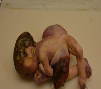
Figure 1: Posterior view of an infant with OEIS complex. Soft tissue mass in the right and lumbosacral lower back/buttock which is covered with skin and represents the myelomeningocele and left omphalocele. Left-equinovarus foot.
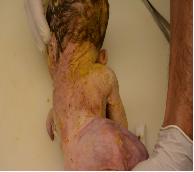
Figure 2: Posterior view of an infant with OEIS complex. Soft tissue mass in the right and lumbosacral lower back/buttock which is covered with skin and represents the myelomeningocele and left omphalocele. Left-equinovarus foot.
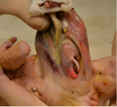
Figure 3: Anterior view of an infant with OEIS complex.
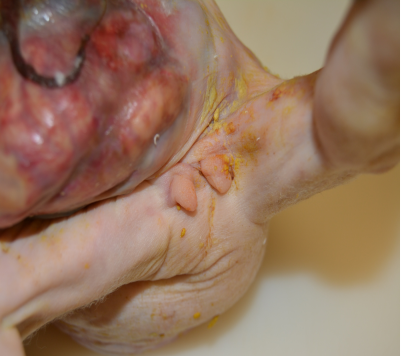
Figure 4: Ambiguous genitalia and lower abdominal wall defect containing abdominal viscera consistent with an omphalocele.
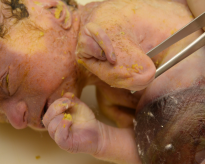
Figure 5: Arthrogryposis involving the left upper limb.
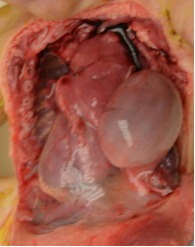
Figure 6: Right-heart
Conclusions
Current study presented the rare finding of coexisting OEIS complex and agenesis of the diaphragm in twin’s pregnancy.
OEIS complex is an entity more frequent in males, being the cases usually sporadic. Some isolated cases in brothers, twin-pregnancy and in chromosomic-related alterations (trisomy in 18, 47, XXX; no-balanced translocation between 9q and Yq) have been described4.
Prenatal diagnostic is possible when there is a defect in the abdominal wall, myelomeningocele and absence of bladder visualisation. Other secondary findings that may drive to prenatal diagnostic: kyphoscoliosis, equinovarus-foot, genitourinary alterations5, single umbilical artery and bladder / spinal defects6.
In physical examination, the case reported here gathered all caudal defects compatible with OEIS Complex. The coexistence with anomalies of upper body like diaphragmatic agenesis, arthrogryposis left arm and right-heart, which cannot be explained by the mechanism typically associated with OEIS Complex, thats why this case is exceptional9.
In almost every case, prognostic is always fatal, depending on the structural defects severity7. An early prenatal diagnostic allows it assessment by a multidisciplinary team, which may allow a more effective genetic advice and management.
Conflict of interest
The authors declare that they have no conflict of interest.
Informed consent
Informed consent was obtained.
References
- Carey JC, Greenbaum B, Hall BD. The OEIS complex (omphalocele exstrophy imperforate anus spinal defects). Birth Defects Orig Artic Ser. 1978; 14(6B): 253-63.
- Kant SG, Bartelings MM, Kibbelaar RE, et al. Severe cardiac defect in a patient with the OEIS complex . Clin Dysmorphol. 1997; 6: 371-374.
- Chen CP. Syndromes and disorders associated with omphalocele (II): OEIS complex and Pentalogy of Cantrell. Taiwan J Obstet Gynecol. 2007; 46: 103-110.
- Comprehensive Genetic Analysis of OEIS Complex Reveals No Evidence for a Recurrent Microdeletion or Duplication. American Journal of Medical Genetics Part A. March 2011; 155A(1): 38-49.
- Anne-Karoline Ebert, Heiko Reutter, Michael Ludwig, et al. The Exstrophy-epispadias complex. Orphanet Journal of Rare Diseases. 2009; volume 4, Article number: 23.
- Kavita Aneja. A rare case of OEIS complex – newer approach to diagnosis of exstrophy bladder by color doppler and its differentiation from simple omphalocele. Indian J Radiol Imaging. 2017 Oct-Dec; 27(4): 436–440.
- Nada Neel, Mohmoud Salem Tarabay. Omphalocele, exstrophy of cloaca, imperforate anus, and spinal defect complex, multiple major reconstructive surgeries needed. Urol Ann. 2018 Jan-Mar; 10(1): 118–121.
- Michael Bamshad, MD, Ann E, Van Heest, MD, et al. Arthrogryposis: A Review and Update. J Bone Joint Surg Am. 2009 Jul 1; 91(Suppl 4): 40–46.
- Kim M. Keppler-Noreuil. OEIS complex (omphalocele-exstrophy-imperforate anus-spinal defects): Areview of 14 cases American Journal of Medical Genetics. 2001; 99: 271±279.